
The Rockwood Classification for AC Joint Separations

The Rockwood Classification for AC Joint Separations
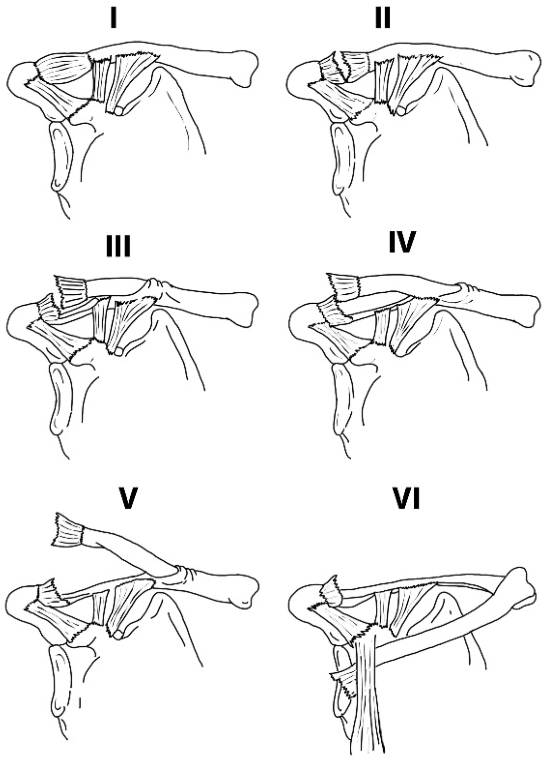
The Rockwood classification is the most widely used system for categorizing acromioclavicular (AC) joint separations, dividing injuries into six types based on ligament disruption, clavicle displacement, and soft tissue involvement.1 Developed in 1984 as an expansion of earlier classifications, it provides a framework for diagnosis and treatment decisions.
Dameron and Rockwood classification (Intact Coracoclavicular ligaments even in high-grade ACJ dislocation).1,2
Type I
- AC ligament: Mild sprain
- Coracoclavicular (CC) ligaments: Intact
- Radiographic findings: Normal appearance with maintained AC joint alignment
- Clinical notes: Minimal pain and swelling, typically treated conservatively
Type II
- AC ligament: Ruptured
- CC ligaments: Sprained but intact
- Radiographic findings: AC joint widening (>7 mm) with <25% vertical clavicle elevation
- Clinical notes: Visible deformity with moderate pain, often managed non-operatively
Type III
- AC ligament: Ruptured
- CC ligaments: Ruptured
- Radiographic findings: CC distance increased 25-100% compared to contralateral side
- Clinical notes: Complete AC joint dislocation with significant deformity; treatment remains controversial
Type IV
- Displacement: Clavicle posteriorly displaced into trapezius muscle
- Soft tissue: Detached deltoid and trapezius muscles
- Radiographic findings: Posterior clavicle position on axillary view
- Notes: Rare injury requiring surgical intervention
Type V
- Displacement: Clavicle subcutaneously elevated
- Radiographic findings: CC distance >100% of contralateral side (>25 mm)
- Soft tissue: Severe detachment of deltotrapezial fascia
- Clinical notes: Dramatic shoulder deformity, typically surgical candidate
Type VI
- Displacement: Clavicle inferior to coracoid process
- Anatomy: Lodged beneath coracobrachialis and biceps tendons
- Notes: Extremely rare, usually from high-energy trauma
The classification relies on standard radiographs (AP, axillary, and weighted views) to assess ligament integrity and displacement patterns. While Types I-II are consistently managed non-operatively and Types IV-VI surgically, Type III remains debated due to variable outcomes with both approaches. Interobserver reliability is moderate, reflecting challenges in radiographic interpretation of soft tissue injuries.
References
- Rockwood CA. Fractures in adults. pp 860–910. Philadelphia: Lippincott, 1984.
- Wahal N, Kendirci AS, Abondano C, Tauber M, Martetschläger F. Acromioclavicular Joint Lesions in Adolescents—A Systematic Review and Treatment Guidelines. Journal of Clinical Medicine. 2023; 12(17):5650. https://doi.org/10.3390/jcm12175650