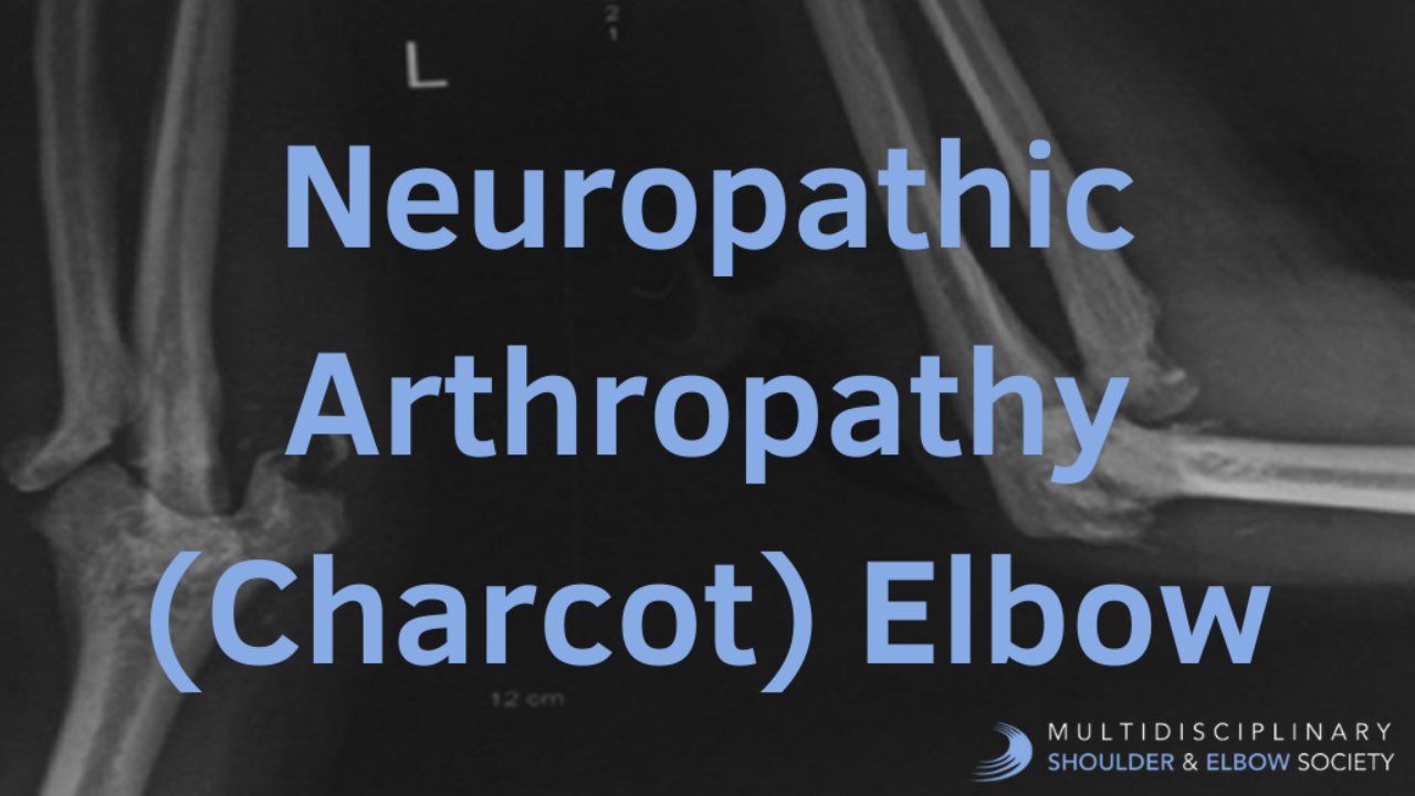
Neuropathic Arthropathy (Charcot) Elbow

Definition
Charcot neuroarthropathy or neuropathic joint or neuropathic arthropathy is a chronic, devastating, and destructive disease of the bone structure and joints in patients with neuropathy; it is characterized by painful or painless bone and joint destruction in limbs that have lost sensory innervation.16 The loss of protective sensation leads to destruction of joints, pathological fractures, surrounding bony structures and debilitating deformities may lead to amputation if left untreated.
History
Jean-Marin Charcot (1825-1893) was a French Neurologist and professor of anatomical pathology. He is known as “the founder of modern neurology.” In 1868 Jean-Martin Charcot gave the first detailed description of the neuropathic joint.17
Epidemiology
The incidence of neuropathic arthropathy is 0.1% to 0.4% of patients with diabetes.17 The prevalence increases to 35% in patients with peripheral neuropathy.18 The risk of developing neuropathic arthropathy is not related to the type of diabetes but more common in people with type I diabetes.19People with Charcot neuroarthropathy are usually in their 50 s or 60 s, and most have had diabetes for at least 10 years. Patients with diabetes and neuropathy have an incidence rate of 7.5%. rare condition in the upper extremity (~ 40 cases reported in literature).
Etiology
Pathophysiology of Neuropathic Arthropathy
Syrinx Formation
Neuropathic arthropathy often begins with syrinx formation, a fluid-filled cavity within the spinal cord that causes damage to the decussating fibers of the lateral spinothalamic tract. This damage leads to dissociative anesthesia, characterized by preserved proprioception and motor function but loss of pain and temperature sensation. As the syrinx enlarges, it extends damage to the dorsal column and anterior horn of the spinal cord, resulting in areflexia, loss of motor strength, and muscle atrophy.1,2
Joint Destruction: Neurotraumatic and Neurovascular Theories
The joint destruction in neuropathic arthropathy occurs through two primary mechanisms. The neurotrauma pathway involves loss of peripheral sensation and proprioception, leading to repetitive microtrauma to the joint. Additionally, poor fine motor control generates unnatural pressure on certain joints, causing further microtrauma.3 The body is unable to adapt a protective mechanism to compensate for microtrauma due to abnormal sensation. The neurovascular pathway involves dysregulated reflexes and desensitized joints that receive significantly greater blood flow. This hyperemia leads to increased osteoclastic resorption of bone.4,5
Genetics
From a molecular biology perspective, the RANK/RANKL/OPG triad pathway is thought to be involved in the pathogenesis of neuropathic arthropathy. This pathway plays a crucial role in bone metabolism and remodeling.6,7
Associated Conditions
Various conditions are associated with neuropathic arthropathy. Orthopedic conditions include ulnar neuropathy, which can lead to joint instability and destruction.8 Among medical conditions and comorbidities, syringomyelia is the most common etiology of neuropathic arthropathy of the upper extremity, accounting for approximately 25% of Charcot joints. Syringomyelia typically causes monoarticular involvement, with the shoulder more commonly affected than the elbow.9,10
The Arnold-Chiari malformation is recognized as the most common cause of syringomyelia.11 Hansen's disease (leprosy) represents the second most common cause of upper extremity neuropathic arthropathy.12 Neurosyphilis, particularly in its tabes dorsalis form, usually affects the knee and can be polyarticular.13 Notably, diabetes remains the most common cause of foot and ankle neuropathic joints in modern clinical practice.14,15
Clinical Presentation
Charcot neuroarthropathy most frequently presents in the 5th decade, with diabetic duration of 20 to 24 years in those with type 2 diabetes. Patients with neuropathic arthropathy commonly present with a swollen elbow. Interestingly, the literature reports that approximately 50% of patients experience pain, while the other 50% present with painless joint destruction.20 Regardless of pain status, loss of function is a predominant complaint among these patients.21 On inspection, the affected joint typically appears swollen, warm, and erythematous, often mimicking an infectious process.22 Various forms of atrophy may be observed due to ulnar nerve entrapment, including interosseous atrophy, hypothenar atrophy, and clawing of the hand.23,24
When examining joint motion, the most common finding is loss of active motion, with elbow flexion, extension, pronation, and supination all potentially affected.25 The elbow joint may demonstrate mechanical instability during examination.26 It's important to note that loss of passive motion specifically indicates a mechanical block within the joint.27
A thorough neurologic evaluation is essential in these cases, as ulnar nerve entrapment at the elbow is very common in patients with neuropathic arthropathy.28 Clinical manifestations of ulnar neuropathy include paresthesias in the ulnar nerve distribution and interosseous weakness.29,30 These neurological findings, combined with the characteristic joint changes, help establish the diagnosis of neuropathic arthropathy.
Imaging Studies
Radiographs
Radiographic evaluation of the elbow typically involves standard views of the affected joint, including anteroposterior (AP) and lateral projections.31
In the early stages of elbow pathology, degenerative changes may mimic osteoarthritis, making diagnosis challenging.31 As the condition progresses, late-stage radiographic findings become more apparent. These include obliteration of the joint space, fragmentation of both articular surfaces leading to subluxation or dislocation, and scattered "chunks" of bone in fibrous tissue. Additional findings may include joint distention by fluid, surrounding soft tissue edema, heterotopic ossification, and fractures. These radiographic changes collectively provide valuable information for assessing the severity and progression of elbow joint pathology.
Magnetic Resonance Imaging
The indication for MRI is to rule out syrinx when neuropathic elbow arthropathy is present.32
Bone Scan
Technetium bone scans
Technetium bone scans may yield positive (hot) results for both neuropathic joints and osteomyelitis, making it challenging to differentiate between these conditions.33
Indium-labeled white blood cell (WBC) scans
Indium-labeled white blood cell (WBC) scans offer a more specific approach to diagnosis. In indium WBC scans, neuropathic joints typically appear negative (cold), while osteomyelitis presents as positive (hot).33 This distinction makes indium WBC scans particularly useful in differentiating between neuropathic joints and osteomyelitis. The combination of these imaging techniques can provide valuable information for accurate diagnosis and appropriate treatment planning in cases where the clinical presentation is ambiguous.33
Studies
Laboratory Studies
The laboratory findings in this condition often include elevated ESR and WBC levels, which can create diagnostic challenges when attempting to differentiate from osteomyelitis.35
Electromyography/Nerve Conduction Study
Electromyography and nerve conduction studies (EMG/NCS) provide valuable diagnostic information, particularly in confirming ulnar neuropathy that is frequently associated with this condition.35
Histology
Histological examination typically reveals characteristic features including synovial hypertrophy and detritic synovitis, which is characterized by the presence of cartilage and bone fragments distributed throughout the synovium.35
Nonoperative Management
Conservative management, including rest, NSAIDs, functional bracing, restriction of activity, and treatment of underlying disease, represents the first line treatment approach for patients with neuropathic elbow joint.36 This non-operative management strategy has demonstrated promising results, with studies indicating that approximately 50% of patients report significant improvement following these interventions (Charcot foot). For cases characterized by severe elbow pain that remains refractory to initial conservative measures, intra-articular corticosteroid injection may be considered as an additional therapeutic option. This targeted intervention can provide relief in patients experiencing intense discomfort and may serve as an intermediary step before considering more invasive surgical approaches in persistent cases.
Operative Management
Neurosurgical decompression is indicated in cases presenting with cervical syrinx, with outcomes demonstrating that decompression of the syrinx has shown to slow disease progression, maximize joint function, and improve bone quality. Studies have documented some elbow joint space restoration following syrinx decompression.37 Peripheral nerve neurolysis is recommended for patients with ulnar nerve palsies or PIN palsies, though limited case series have shown good recovery of nerve function but with high recurrence rates. Elbow arthrodesis becomes a consideration for patients experiencing elbow pain and instability who have failed conservative management; however, limited case series have shown improvement of pain but with accompanying functional limitations.38 Regarding total joint replacement, it should be noted that Charcot joint is considered a contraindication to elbow total joint replacement due to poor bone stock, prosthetic loosening, instability, and soft-tissue compromise. The literature contains limited case reports on elbow arthroplasties for Charcot elbow, with these studies showing mixed results.39
References
-
Nogués MA, Roncoroni AJ, Plencovich M. Syringomyelia as a cause of neurogenic arthropathy. Medicina (B Aires). 2017;77(2):141-144.
-
Tubbs RS, Demerdash A, Vahedi P, et al. Clinical anatomy of the craniovertebral junction and cervical spine. Clin Anat. 2015;28(3):345-351.
-
Rogers LC, Frykberg RG. The Charcot foot. Med Clin North Am. 2013;97(5):847-856.
-
Jeffcoate WJ, Game F, Cavanagh PR. The role of proinflammatory cytokines in the cause of neuropathic osteoarthropathy (acute Charcot foot) in diabetes. Lancet. 2005;366(9502):2058-2061.
-
La Fontaine J, Lavery L, Jude E. Current concepts of Charcot foot in diabetic patients. Foot (Edinb). 2011;21(4):187-194.
-
Mabilleau G, Petrova NL, Edmonds ME, et al. Increased osteoclastic activity in acute Charcot's osteoarthropathy: the role of receptor activator of nuclear factor-kappaB ligand. Diabetologia. 2011;54(4):832-841.
-
Petrova NL, Edmonds ME. A prospective study of calcaneal bone mineral density in acute Charcot osteoarthropathy. Diabetes Care. 2016;29(9):2190-2194.
-
Breen TF, Jupiter JB, Searles JS, et al. Neuropathic arthropathy of the elbow in syringomyelia: a report of five cases. J Bone Joint Surg Am. 2013;95(17):e1201-e1206.
-
Hatzis N, Kaar SG, Wirth MA, et al. Neuropathic arthropathy of the shoulder. J Bone Joint Surg Am. 2014;80(9):1314-1319.
-
Atalar AC, Sungur M, Demirhan M, et al. Neuropathic arthropathy of the shoulder associated with syringomyelia: a report of six cases. J Bone Joint Surg Br. 2010;92(1):112-116.
-
Tubbs RS, Oakes WJ. The Chiari malformations: a historical context. In: The Chiari Malformations. Springer; 2004:5-11.
-
Spiegel DA, Singh GK, Banskota AK. Tuberculosis of the musculoskeletal system. Tech Orthop. 2005;20(2):167-178.
-
Allali F, Rahmouni R, Hajjaj-Hassouni N. Tabetic arthropathy. A report of 43 cases. Clin Rheumatol. 2006;25(6):858-860.
-
Wukich DK, Sung W. Charcot arthropathy of the foot and ankle: modern concepts and management review. J Diabetes Complications. 2009;23(6):409-426.
-
Rogers LC, Frykberg RG, Armstrong DG, et al. The Charcot foot in diabetes. Diabetes Care. 2011;34(9):2123-2129.
-
Edmonds ME. Progress in care of the diabetic foot. Lancet. 1999;354(9175):270-272. doi:10.1016/s0140-6736(99)90012-0
-
Dardari D. An overview of Charcot's neuroarthropathy. J Clin Transl Endocrinol. 2020 Oct 28;22:100239. doi: 10.1016/j.jcte.2020.100239. PMID: 33251117; PMCID: PMC7677697.
-
Schoots IG, Slim FJ, Busch-Westbroek TE, Maas M. Neuro-osteoarthropathy of the foot-radiologist: friend or foe?. Semin Musculoskelet Radiol. 2010;14(3):365-376. doi:10.1055/s-0030-1254525
-
Petrova NL, Edmonds ME. Acute Charcot neuro-osteoarthropathy. Diabetes Metab Res Rev. 2016;32 Suppl 1:281-286. doi:10.1002/dmrr.2734
-
Minami A, Ogino T, Sakuma T, Usui M. Neuropathic arthropathy of the elbow. J Bone Joint Surg Br. 1989;71(2):277-281.
-
Ruette P, Stuyck J, Debeer P. Neuropathic arthropathy of the shoulder and elbow associated with syringomyelia: a report of 3 cases. Acta Orthop Belg. 2007;73(4):525-529.
-
Nacir B, Cebeci SA, Cetinkaya E, et al. Neuropathic arthropathy progressing with multiple joint involvement in the upper extremity due to syringomyelia and type I Arnold-Chiari malformation. Rheumatol Int. 2010;30(7):979-983.
-
Eichenbaum MD, Brems JJ. Neuropathic elbow arthropathy: treatment with total elbow arthroplasty. J Shoulder Elbow Surg. 2009;18(6):886-890.
-
Millender LH, Nalebuff EA, Holdsworth DE. Posterior interosseous-branch compression syndrome secondary to rheumatoid synovitis. J Bone Joint Surg Am. 1973;55(4):753-757.
-
Hagert E, Ljung BO, Forsgren S. General innervation pattern and sensory corpuscles in the scapholunate interosseous ligament. Cells Tissues Organs. 2004;177(1):47-54.
-
Deirmengian CA, Lee SG, Jupiter JB. Neuropathic arthropathy of the elbow: a report of five cases. J Bone Joint Surg Am. 2001;83-A(6):839-844.
-
Karthik K, Nanda R, Stothard J. Charcot elbow—a case report. J Shoulder Elbow Surg. 2008;17(4):e1-e3.
-
Culp RW, Osterman AL, Davidson RS, et al. Neural injuries associated with supracondylar fractures of the humerus in children. J Bone Joint Surg Am. 1990;72(8):1211-1215.
-
Rawal N, Faust F, Bryan BM 3rd, et al. Neuropathic shoulder arthropathy secondary to syringomyelia. J Surg Orthop Adv. 2007;16(4):183-186.
-
Yanik B, Tunç H, Şahin B. Neuropathic arthropathy caused by Arnold-Chiari malformation with syringomyelia. Rheumatol Int. 2004;24(4):238-241.
-
Snoddy MC, Lee DH, Kuhn JE. Charcot shoulder and elbow: a review of the literature and update on treatment. J Shoulder Elbow Surg. 2017;26(3):544-552. doi:10.1016/j.jse.2016.10.015
-
Deng X, Wu L, Yang C, Xu Y. Neuropathic arthropathy caused by syringomyelia. J Neurosurg Spine. 2013;18(3):303-309. doi:10.3171/2012.11.SPINE12860
-
Palestro CJ, Mehta HH, Patel M, et al. Marrow versus infection in the Charcot joint: indium-111 leukocyte and technetium-99m sulfur colloid scintigraphy. J Nucl Med. 1998;39(2):346-350.
-
Saltzman CL, Hagy ML, Zimmerman B, Estin M, Cooper R. How effective is intensive nonoperative initial treatment of patients with diabetes and Charcot arthropathy of the feet?. Clin Orthop Relat Res. 2005;(435):185-190. doi:10.1097/00003086-200506000-00026
-
Vopat ML, Nentwig MJ, Chong ACM, Agan JL, Shields NN, Yang SY. Initial Diagnosis and Management for Acute Charcot Neuroarthropathy. Kans J Med. 2018 Nov 29;11(4):114-119. PMID: 30937152; PMCID: PMC6276967.
-
Waibel FWA, Böni T. Nonoperative Treatment of Charcot Neuro-osteoarthropathy. Foot Ankle Clin. 2022;27(3):595-616. doi:10.1016/j.fcl.2022.05.002
-
Deng X, Wu L, Yang C, Xu Y. Neuropathic arthropathy caused by syringomyelia. J Neurosurg Spine. 2013;18(3):303-309. doi:10.3171/2012.11.SPINE12860
-
Jen CL, Tan JC. Neuropathic arthropathy of the elbow treated with double-plate arthrodesis and resection site bone graft. Shoulder Elbow. 2016 Jan;8(1):48-53. doi: 10.1177/1758573215598731. Epub 2015 Aug 11. PMID: 27583001; PMCID: PMC4935168.
-
Nakamura H, Iwamoto T, Kimura H, et al. A case of neuropathic arthropathy of the elbow with early loosening after total elbow arthroplasty. Mod Rheumatol Case Rep. 2023;7(2):480-482. doi:10.1093/mrcr/rxac094
