
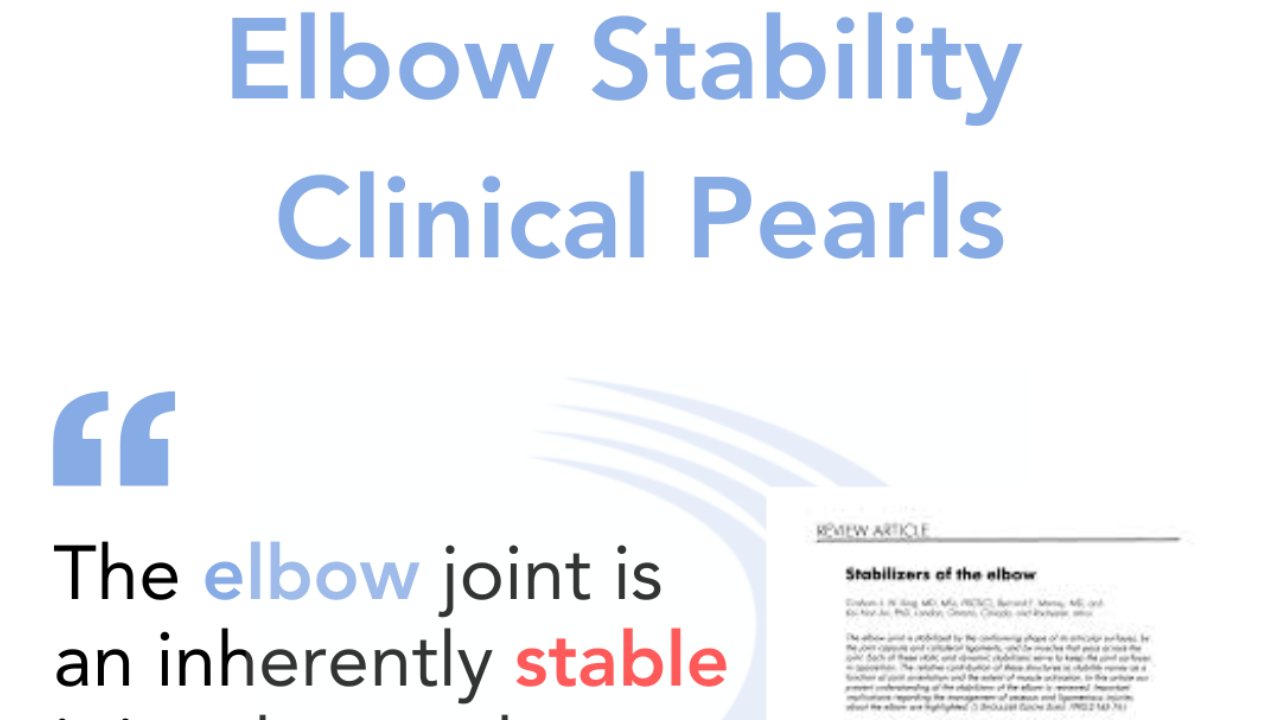
Elbow Stability Clinical Pearls
Centerline of Rotation
The elbow's centerline of rotation is created by the trochlea-trochlear notch articulation12. It's located 16.54 mm from the joint line towards the medial epicondyle along the anterior bundle of the medial ulnar collateral ligament (AMCL)2. This axis isn't constant throughout flexion-extension motion13.
Soft Tissue Stabilizers
The joint capsule and collateral ligaments provide static constraint. The origin of the lateral collateral ligament complex on the lateral epicondyle is near the axis of rotation. It remains taut throughout the flexion extension arc of elbow motion. The lateral ulnar collateral ligament (LUCL) originates on the lateral epicondyle and inserts on the cristae supinatoris of the ulna, and is an important stabilizer in both rotation and varus stability. The anterior band of the medial collateral ligament (MCL) originates on the medial epicondyle and inserts on the sublime tubercle of the ulna and is the primary valgus stabilizer of the elbow.
Bony Static Stabilizers
The elbow joint is inherently stable due to the interlocking shape of its articular surfaces. Elbow stability involves three articulations: humeroulnar, radiocapitellar, and proximal radioulnar4. Bony interactions are more important in full extension and flexion, less so between 30° to 100° of flexion4.
Ulnohumeral Articulation
The ulnohumeral joint is the primary bony stabilizer4. The coronoid process is crucial for stability, with 50% articular surface removal as the cutoff for instability4.
Radiocapitellar Articulation
The radial head facilitates flexion-extension and rotation, transmits forces, and provides tension to the lateral ulnar collateral ligament4. It's a secondary valgus stability restraint4.
Dynamic Stabilizers
Muscles originating from the humerus and forearm provide dynamic stability4. The flexor-pronator mass assists the AMCL in resisting valgus stress4. Forearm muscles can also resist varus forces4.
Biomechanical Importance
Different regions of the elbow have varying biomechanical importance. The coronoid is a primary stabilizer, while the proximal olecranon can be partly resected without substantial kinematic alterations4.
References
- Graham KS, Golla S, Gehrmann SV, Kaufmann RA. Quantifying the Center of Elbow Rotation: Implications for Medial Collateral Ligament Reconstruction. Hand (N Y). 2019;14(3):402-407. doi:10.1177/1558944717743599
- Liu, H., Kholinne, E., Sun, Y. et al. The dynamic rotation axis of ulnohumeral joint during active flexion-extension: an in vivo 4-dimensional computed tomography analysis. BMC Musculoskelet Disord 23, 152 (2022). https://doi.org/10.1186/s12891-022-05102-5
- Wilps T, Kaufmann RA, Yamakawa S, Fowler JR. Elbow Biomechanics: Bony and Dynamic Stabilizers. J Hand Surg Am. 2020;45(6):528-535. doi:10.1016/j.jhsa.2020.01.016
- Karbach LE, Elfar J. Elbow Instability: Anatomy, Biomechanics, Diagnostic Maneuvers, and Testing. J Hand Surg Am. 2017 Feb;42(2):118-126. doi: 10.1016/j.jhsa.2016.11.025. PMID: 28160902; PMCID: PMC5821063.